
How 3D Imaging Changes The Way Oral Surgeons Plan Complex Procedures
Some oral surgery procedures require more than a standard X-ray can show. When a case involves impacted teeth, limited bone, nearby nerves, the sinuses, or a planned dental implant, your oral surgeon may recommend 3D imaging to get a more detailed view of the area. A 3D scan can show the height, width, depth, and position of these structures from multiple angles, giving your surgeon more information to consider when planning treatment.
3D imaging does not replace your consultation or clinical examination. Instead, it adds another layer of information that can help your surgeon understand your anatomy and determine which treatment options may be appropriate.
In this blog, we’ll explain how 3D imaging works, what a dental CT scan and cone beam imaging can show, when they’re useful, and how they can support treatment planning for implants, wisdom teeth, bone grafting, and other complex procedures.
From A Flat X-Ray To A 3D Surgical Map
Think of a 2D X-ray as a photograph. It gives a useful picture, but the image is still flat. A 3D scan is more like a map. It lets the surgeon examine height, depth, width, and position from different angles.
That extra information can matter when a tooth is impacted, bone is thin, a nerve is nearby, or the sinus is close to an implant site. A dental CT scan can give the surgeon a fuller picture before treatment begins.
What A Dental CT Scan Can Show
A dental CT scan may show bone height and width, root positions, impacted teeth, jaw shape, infection patterns, nerve pathways, sinus position, and changes that may not be obvious on a standard X-ray.
You do not need to read the scan yourself. The surgeon reviews the images along with your exam and medical history, then explains what they mean and whether another step may be needed. This is how 3D imaging in oral surgery can make treatment planning more informed.
Cone Beam Imaging: The Technology Behind The View
Cone beam imaging, often called CBCT, is a type of 3D dental imaging used in oral and maxillofacial surgery. The scanner rotates around your head to create a three-dimensional view of the area being examined, allowing your surgeon to review the structures from different angles.
The scan is generally quick and noninvasive, and you may sit or stand while the images are captured. Like any diagnostic tool, CBCT is not needed for every patient. Your surgeon may recommend it when additional detail could help evaluate your anatomy or plan your treatment.
Why 3D Detail Matters For Dental Implants
Dental implants require careful planning. Your surgeon must consider bone volume, nearby teeth, nerves, sinuses, and the intended implant position. When bone is limited, treatment may include grafting, ridge preservation, or a sinus lift.
With 3D imaging and oral surgery planning, the surgeon can assess whether enough bone is present and where the implant may fit best. A dental CT scan can also show nearby structures that need consideration.
This additional detail can help your surgeon understand your anatomy before treatment and build a plan around your specific needs. The goal is not simply to use more technology, but to have the right information available when precise planning matters.
Wisdom Teeth, Impacted Teeth, And Hidden Anatomy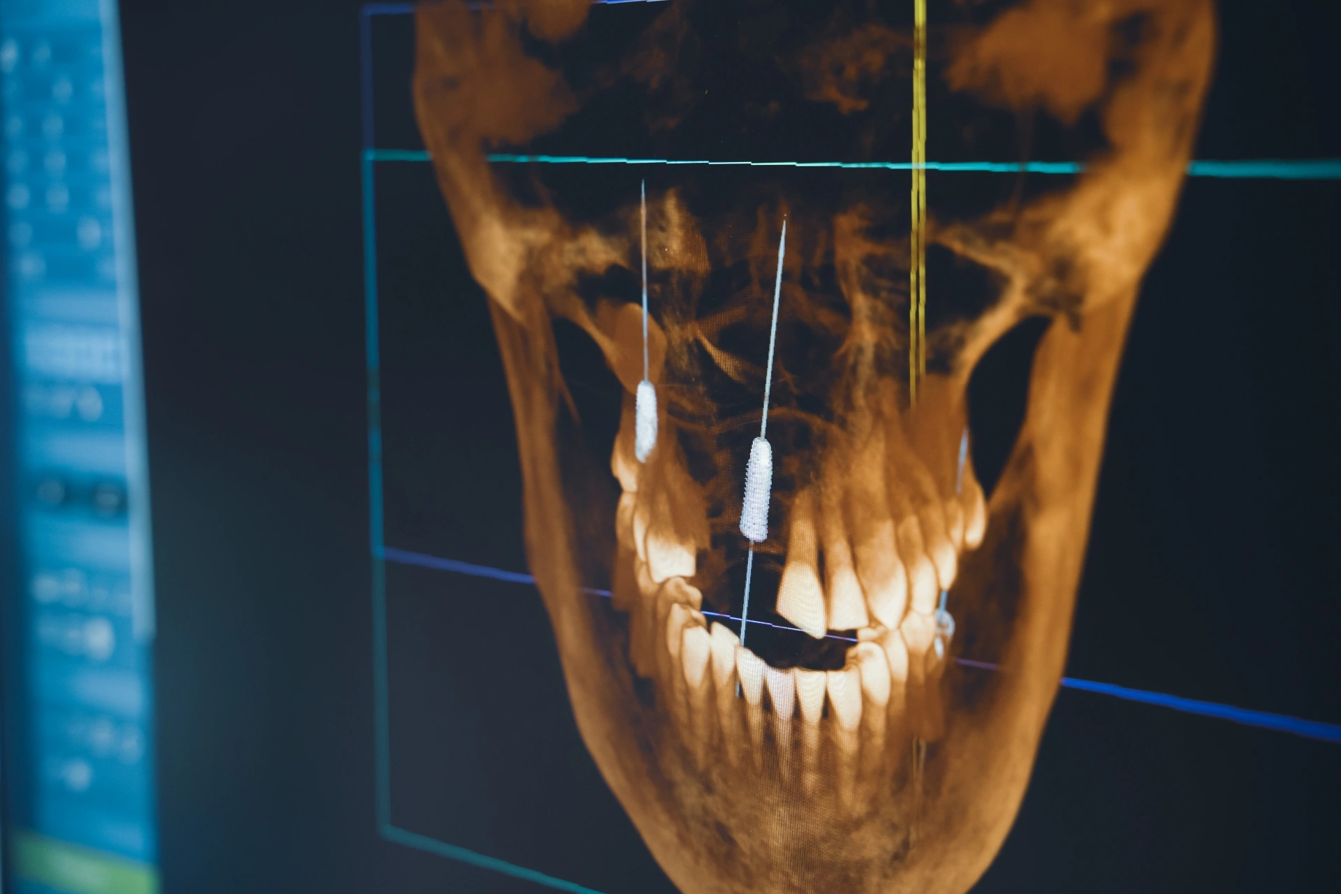
Impacted teeth can develop at different angles, remain partially or fully enclosed in bone, or sit close to nearby roots and nerves. In these situations, a standard X-ray may not provide enough detail to fully understand the tooth’s position and its relationship to surrounding structures.
A 3D CT scan provides a more detailed view of the tooth from different angles, helping your surgeon assess its position and surrounding anatomy before treatment. However, 3D imaging is not necessary for every impacted wisdom tooth. Your surgeon can determine whether the additional imaging is appropriate based on your examination and the complexity of the case.
Bone Grafting And Sinus Lift Planning
Bone grafting and sinus lift procedures also require proper planning. The surgeon needs to know how much bone is present and whether any additional support is required.
3D imaging for oral surgery planning can help assess bone height, bone width, sinus location, and grafting needs before the procedure. This can be especially helpful when preparing the upper jaw for dental implants.
Oral Pathology And Unusual Findings
Sometimes an oral surgeon needs to examine a cyst, lesion, swelling, or other unusual finding more closely. Advanced oral surgery technology may help show its size, location, and relationship to nearby structures.
The scan is only one part of the evaluation. Symptoms, the clinical exam, dental history, and medical history also matter. Depending on the findings, the next step could be monitoring, a biopsy, or treatment.
What Patients Can Expect During the Scan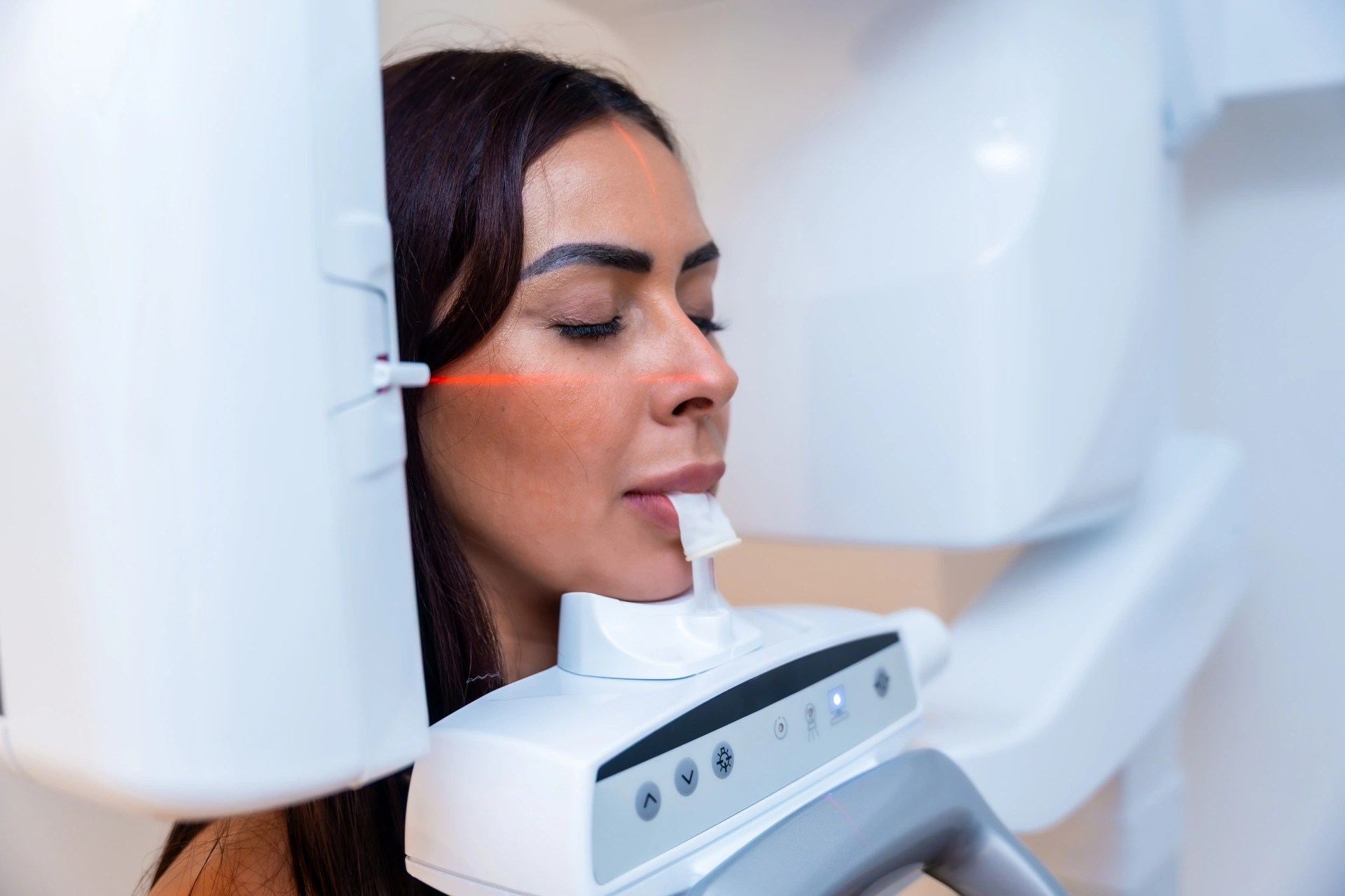
A dental CBCT scan usually requires little preparation and is generally quick and painless. Depending on the scanner, you may sit or stand while your head is positioned, and you will need to remain still while the images are captured. You may be asked to remove jewelry, glasses, or removable dental appliances that could interfere with the scan.
Once the scan is complete, your oral surgeon can review the images alongside your examination and medical history to help plan your treatment. Since CBCT uses X-rays, it should be recommended when the additional information is clinically useful, not as a routine step for every patient.
If you are pregnant or think you may be pregnant, let your dental team know before the scan so they can determine whether the imaging is necessary and appropriate
Why More Information Can Lead To A Personalized Plan
Every patient has different anatomy. One person may have enough bone for an implant right away, while another may need grafting. One impacted tooth may be straightforward, while another may require closer planning.
That is where oral surgery technology can be useful. The goal is not to use more technology simply because it is available. It is to use the right information to create a treatment plan that fits the patient.
FAQs
What is 3D imaging oral surgery planning?
Three-dimensional imaging planning for oral surgery uses advanced imaging techniques to visualize teeth, bone, nerves, sinuses, and adjacent structures before surgery.
Is a dental CT scan the same as a regular X-ray?
No. Dental X-rays give you a two-dimensional picture, while a CT scan gives you a three-dimensional view
What is cone beam imaging?
Cone beam imaging, or CBCT, is a type of 3D dental scan that shows oral and facial structures from multiple angles.
Why would an oral surgeon use 3D imaging before dental implants?
It may help evaluate bone volume, nerve location, sinus position, and possible implant placement options.
Is 3D imaging needed for wisdom teeth removal?
Sometimes. It may be recommended when a wisdom tooth is close to a nerve, deeply impacted, or difficult to assess on a standard X-ray.
Does 3D imaging replace the consultation?
No. Imaging supports the consultation. Your surgeon also considers your symptoms, health history, exam findings, and treatment goals.
Better Planning Starts Here
A 3D scan does not replace your oral surgeon’s examination or clinical judgment. Instead, it provides additional information when a case requires more detailed planning. For dental implants, impacted teeth, wisdom teeth, bone grafting, sinus lifts, and certain pathology concerns, this added detail can help your surgeon better understand your anatomy and discuss appropriate treatment options.
At Premier Oral, we use 3D imaging when it supports thoughtful, personalized care. Schedule a consultation with us to discuss your treatment and learn whether 3D imaging may be appropriate for your needs.
Comments are closed.